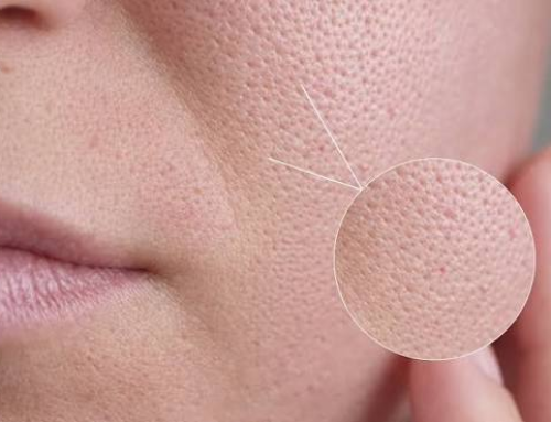
 Most patients who come in asking how to remove white patches on the face have already tried something — a cream recommended by a pharmacist, a home remedy, sometimes a steroid ointment left over from an old prescription. Very few of these attempts work, and the reason is rarely the product. So if you’re wondering how to remove white patches on the face, the first step is understanding what is causing them. White patches are not a single disease—they are a symptom with several possible causes, and each requires a different treatment and a product that helps one can do nothing at all for another, or occasionally make it worse. Getting rid of white patches on the face reliably starts with identifying which of these is actually causing them.
What Actually Causes White Patches on the Face
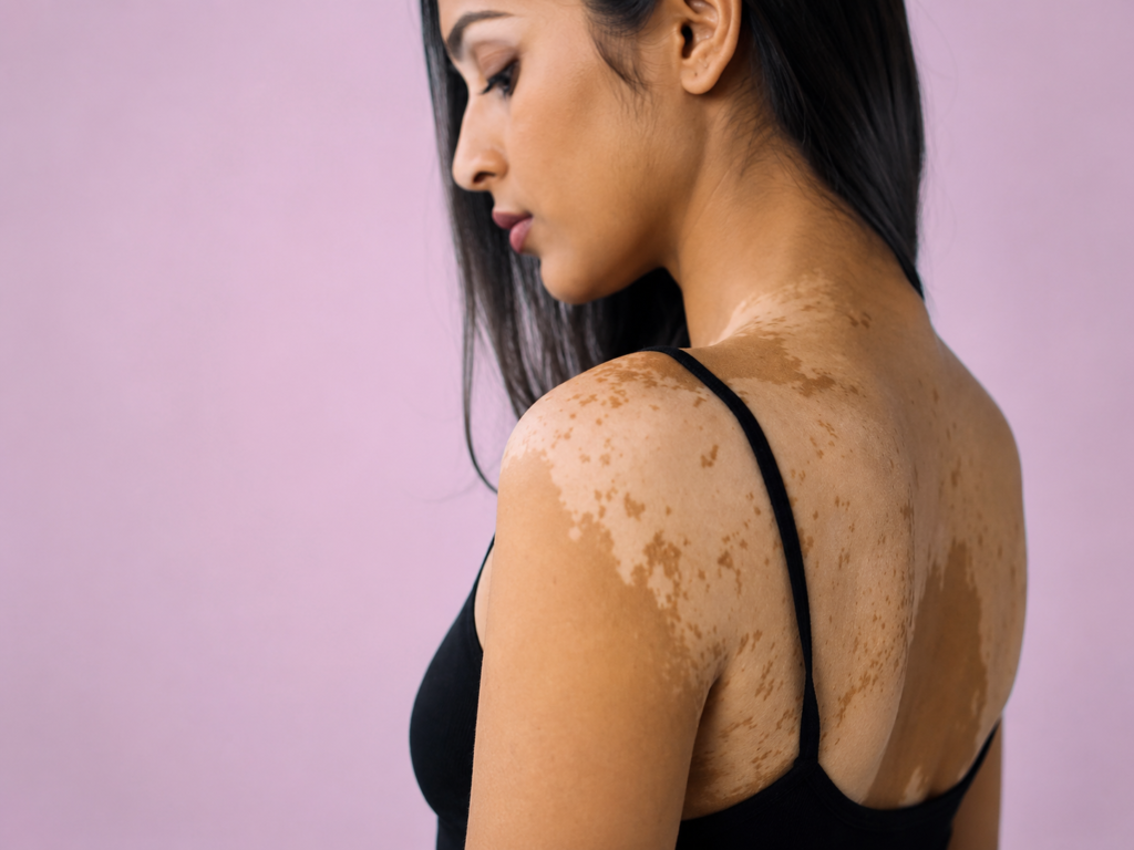
Vitiligo is an autoimmune condition in which the immune system destroys melanocytes, the pigment-producing cells in skin. Patches are typically well-demarcated, milky-white, and can enlarge or appear symmetrically on both sides of the face. Hair within a vitiligo patch can also turn white — a detail that helps distinguish it from other causes. Pityriasis alba is a mild, eczema-related condition, most common in children and teenagers. It produces ill-defined, faintly scaly, pale patches — usually on the cheeks — that are more noticeable after sun exposure because the surrounding skin tans while the patch does not. It is not a loss of pigment cells; it is a temporary under-function of the pigment they already have. 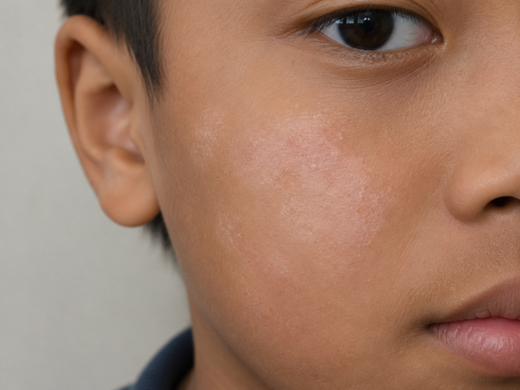 Tinea versicolor, caused by an overgrowth of Malassezia yeast that is naturally present on skin, can present as pale, finely scaled patches on the face as well as the trunk. It is fungal, not autoimmune, and is covered in more detail in Tinea Versicolor — The Fungal Infection Causing Uneven Skin Tone. Post-inflammatory hypopigmentation follows an injury to the skin — healed acne, eczema, a burn, or an aggressive peel or laser treatment. The inflammation temporarily suppresses melanin production at the site, leaving a pale patch that usually — though not always — recovers on its own over months. Idiopathic guttate hypomelanosis is less common on the face than on sun-exposed arms and shins, but does occasionally appear on the forehead and temples in adults past their thirties. It produces small, sharply defined white spots, typically a few millimetres wide, linked to cumulative sun exposure rather than any autoimmune or infectious process. Unlike the other causes on this list, it does not spread or enlarge — it simply accumulates as more spots over years.
Why “Removing” Isn’t the Right Framing
A white patch is an area with less pigment than the skin around it, not an area with something extra sitting on top of it. There is nothing to remove. The real clinical question is why melanocytes at that spot have stopped producing pigment, or have been destroyed altogether and the answer determines whether the patch can be brought back to normal colour, needs to be prevented from spreading, or will resolve without intervention. 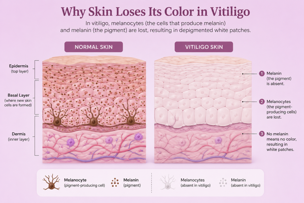 Treating a pigment-loss condition with an exfoliant or a brightening serum, as many patients do before their first consultation, addresses none of these mechanisms. This is also why fairness creams, home remedies involving lemon or turmeric, and over-the-counter depigmentation products consistently fail on these patches specifically — even when they visibly work on the surrounding normal skin. Those products act on melanin already present in functioning melanocytes. In a true white patch, that mechanism does not exist to act on.
How the Diagnosis Is Actually Made
The distribution, border quality, scale, and any concomitant symptoms (itching in pityriasis alba, minor flaking in tinea versicolor) immediately narrow the diagnosis, thus a clinical examination is usually sufficient. A Wood’s lamp, which fluoresces under UV light and makes vitiligo’s borders much more distinct than they appear in normal light, and a potassium hydroxide (KOH) skin scraping, which confirms or rules out a fungal cause like tinea versicolor within minutes, are two easy in-clinic tools that help when the picture is unclear. A biopsy is rarely required, and it should only be used for unusual presentations that don’t fit the conventional patterns. Laser hair reduction can be considered as a treatment option in suitable cases.
White Patches on Face Treatment: What Works for Each Cause
- Vitiligo: For face, topical corticosteroids or calcineurin inhibitors (tacrolimus 0.1%) are the first line of treatment; for larger patches, they are sometimes paired with narrowband UVB (NB-UVB) phototherapy. It usually takes three to six months of constant treatment to see obvious repigmentation, and the earlier treatment begins, the better the results are.
- Pityriasis alba: A short course of a mild topical steroid to settle the underlying inflammation, followed by consistent moisturisation and daily sunscreen. The contrast fades as the surrounding tan evens out; most cases resolve on their own within months to a couple of years.
- Tinea versicolor: Topical or oral antifungals clear the yeast overgrowth, but pigment normalisation lags behind — patches can take several weeks to months to even out even after the fungus is gone.
- Post-inflammatory hypopigmentation: Largely a matter of patience and sun protection. Sunscreen prevents the surrounding skin from tanning further and exaggerating the contrast. In persistent cases, procedures such as targeted phototherapy can accelerate repigmentation.
- Idiopathic guttate hypomelanosis: Not reversible with topical treatment, since the melanocytes at each spot are reduced rather than temporarily suppressed. Options are largely cosmetic — targeted micro-needling or fractional laser to stimulate some pigment return in individual spots — combined with consistent sun protection to prevent new ones from forming.
| Cause | Appearance | Treatment |
| Vitiligo | Milky-white, well-demarcated, can enlarge; hair within patch may whiten | Topical corticosteroids/tacrolimus 0.1%, NB-UVB phototherapy |
| Pityriasis alba | Ill-defined, faintly scaly, pale; more visible after tanning | Mild topical steroid, moisturiser, daily sunscreen |
| Tinea versicolor | Pale, finely scaled patches, sometimes itchy | Topical or oral antifungals |
| Post-inflammatory hypopigmentation | Pale patch at site of prior injury or inflammation | Sun protection and time; phototherapy if persistent |
| Idiopathic guttate hypomelanosis | Small (few mm), sharply defined white spots; doesn’t spread | Micro-needling or fractional laser, sun protection |
What You Can Do While Waiting for Treatment to Work
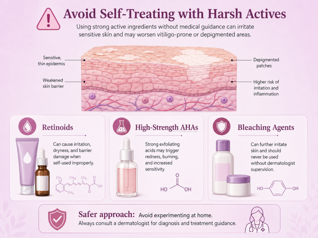 None of these conditions respond overnight, and the single most useful thing a patient can do in the interim is daily sunscreen — broad-spectrum, SPF 30 or higher, reapplied through the day. Untreated sun exposure tans the surrounding skin further, which widens the visible contrast even if the patch itself is not changing size. It also means that self-treatment with harsh actives — retinoids, high-strength AHAs, or bleaching agents — should be paused, since irritating the area around a white patch can worsen post-inflammatory hypopigmentation and, in vitiligo, has been observed to occasionally trigger new patches at sites of skin trauma (a phenomenon called the Koebner response). Camouflage make-up formulated for pigment mismatch is a reasonable short-term option while medical treatment takes effect — it changes nothing about the underlying condition, but it is not harmful either. Chemical peels may also be considered as a treatment option in suitable cases.
When to See a Dermatologist
A white patch that is spreading, sharply bordered, symmetric, or accompanied by whitening of the hair within it warrants an evaluation for vitiligo specifically, since early treatment materially changes the outcome. More broadly, because these conditions can look similar to the eye but require entirely different treatments, an accurate diagnosis — usually possible on clinical examination, sometimes confirmed with a Wood’s lamp or a simple skin scraping — should come before any white spots on face treatment or white patches on skin treatment is started.
Frequently Asked Questions
Are white patches on the face contagious?
No. Vitiligo, pityriasis alba, and post-inflammatory hypopigmentation are not caused by anything transmissible. Tinea versicolor is fungal, but the Malassezia yeast behind it is already present on everyone’s skin — the patches come from local overgrowth, not transmission from another person.
Can white patches on the face be completely cured?
It depends on the cause. Pityriasis alba and post-inflammatory hypopigmentation typically resolve fully on their own or with treatment, and tinea versicolor clears with antifungals, though pigment takes weeks to normalise afterward. Vitiligo is a chronic autoimmune condition — repigmentation is achievable for many patients with sustained treatment, but it is managed rather than cured outright, and recurrence is possible.
Do white patches on the face spread if left untreated?
Vitiligo can enlarge or produce new patches, particularly during active phases, which is why early evaluation matters. The other causes here — pityriasis alba, tinea versicolor, post-inflammatory hypopigmentation, idiopathic guttate hypomelanosis — do not typically spread the same way, though tinea versicolor can extend locally if the underlying yeast overgrowth goes untreated.
Can a vitamin deficiency cause white patches on the face?
Deficiencies in vitamin B12, folate, or copper have been linked to pigment changes in some studies, but they are an uncommon cause of well-defined white patches and shouldn’t be assumed as the explanation without a proper diagnosis. Correcting a genuine deficiency can help in the rare cases where it contributes, but it will not resolve vitiligo, pityriasis alba, or a fungal cause.
Does stress cause white patches on the face?
Stress doesn’t directly cause any of these conditions, but it is recognised as a possible trigger for new vitiligo patches or flare-ups in people who already carry the underlying autoimmune predisposition. It is not a standalone cause, and it plays no role in pityriasis alba, tinea versicolor, or post-inflammatory hypopigmentation.
Is a white patch on the face always vitiligo?
No — vitiligo is one of several possible causes, not a synonym for the symptom itself. Assuming every white patch is vitiligo, or dismissing vitiligo because a patch looks minor, are both common misreadings. Border quality, hair involvement, scale, and distribution are what actually tell them apart. At our clinic, diagnosis is usually confirmed through clinical examination and, where required, Wood’s lamp evaluation or KOH microscopy before recommending treatment.
Book a consultation: wa.me/919381218003
— Dr. Nishita Ranka | Consultant Dermatologist | Dr. Nishita’s Clinic for Skin, Hair & Aesthetics, Hyderabad